Research projects
In our group we study the structure and function of VWA domain-containing extracellular matrix proteins. We recombinantly express full-length proteins and protein fragments in prokaryotic and eukaryotic systems. The proteins are not only used for structural and functional studies, but also to generate highly specific antibodies, which allow us to perform detailed tissue distribution studies. CD spectroscopy is used to determine the general folding and thermal stability of the recombinant proteins.
Interactions are studied by ELISA style binding assays and surface plasmon resonance spectroscopy to gain insight into the supramolecular assembly of the proteins in the extracellular matrix. The structure of particular domains is revealed by NMR, SAXS and X-ray crystallography. By site directed mutagenesis we study the consequences of disease causing mutations at the cellular and molecular level.
We gain insight into the biological functions of the proteins we study through "knockout" experiments in mouse as well as transient "knockdowns" in the developing zebrafish. These investigations will lead to an increased understanding of the functions of a particular subgroup of extracellular matrix proteins and their involvement in the pathogenesis of certain diseases like Multiple Epiphyseal Dysplasia (MED) or collagen VI related muscular dystrophies.
Collagen VI
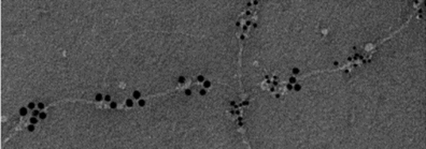
Collagen VI microfibrils are widespread in the extracellular matrix (EM) of musculoskeletal tissues and connect large interstitial structures and cells. They are affected by mutations in Bethlem Myopathy and Ullrich Congenital Muscular Dystrophy and besides collagen VI is upregulated in osteoarthritis. Further, a 7kDa fragment referred to as “endotrophin” was recently proposed to act as an adipokine enhancing tumor progression, fibrosis, inflammation and insulin resistance. The structure of collagen VI is unusual. The polypeptide chains are composed of only a short triple helix and predominant tandem arrays of globular von Willebrand factor type A (VWA) domains that often bind ligands in a metal ion dependent manner. Three different chains assemble intra- and extracellularly by a complex series of chain interactions and proteolytic cleavages that are only partially understood. The secreted form is a tetramer of heterotrimeric monomers and has a molecular mass of about 2,200 kDa. Fully assembled microfibrils also carry other proteins that are covalently or non-covalently linked. Our goal is to elucidate the interactions, assembly and degradation of collagen VI to reveal pathomechanisms in musculoskeletal disorders, e.g. myopathies and osteoarthritis. This would potentially allow the development of novel therapeutic approaches. We express single or tandem domains of collagen VI in both bacteria and eukaryotic cells at large scale to set up crystal trays and perform SAXS measurements to localize interaction surfaces for interchain assembly. We focus on the ligand binding VWA domains and on the determination of proteolytic cleavage sites. A major critical step in the intracellular assembly is the dimerization of two heterotrimeric monomers by an interaction between a VWA domain and a particular triple helical region. Indeed, mutations leading to severe myopathy phenotypes accumulate in this region. Patient-derived fibroblast cell lines carrying mutations in collagen VI chains or immortalized fibroblast and chondrogenic cell lines manipulated by CRISPR-Cas will be used to define the steps in the assembly when VWA domain interactions or proteolytic cleavage trigger pathomechanisms in muscle and cartilage. We will perform immunohistochemistry on cell cultures and western blots on cell lysates and supernatants using our unique array of antibodies against all N- and C-terminal VWA domain containing globular regions and against the endotrophin fragment to analyze interactions, assembly and proteolytic fragmentation. Finally, the determination of crucial interaction sites and cleavage sites will allow us to generate mouse strains by CRISPR-Cas where these sites have been mutated to study pathomechanisms of musculoskeletal disorders in vivo.
AMACO
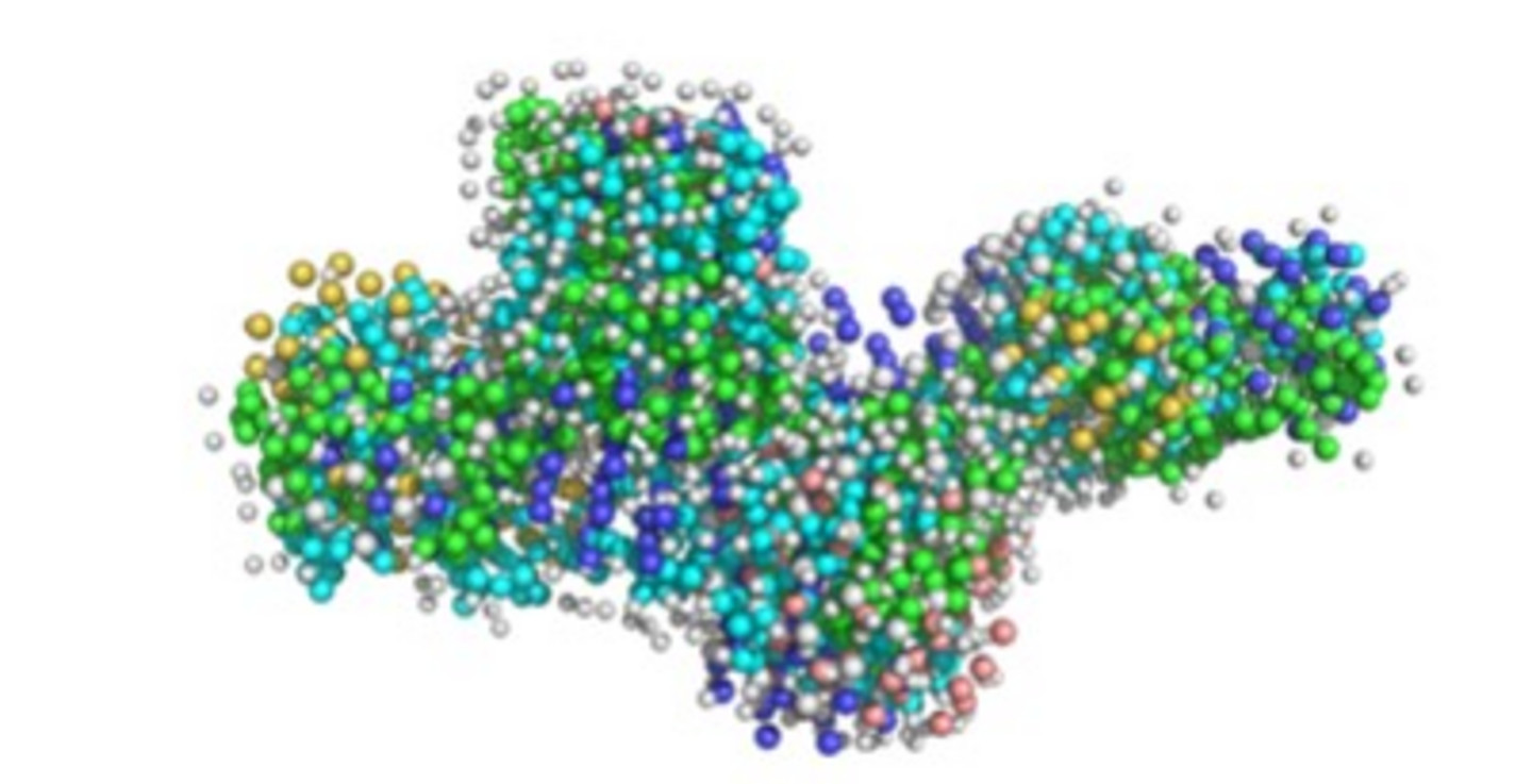
Extracellular microfibrillar and filamentous suprastructures control tissue integrity and cell fate. We study the physical and/or functional interaction between the Fraser complex and the novel AMACO-containing suprastructure. We will investigate the organization of the AMACO-containing suprastructure, its cellular receptors and its integration into the Fraser complex. The in vivo functions of AMACO are studied in a mouse model. Tumor samples are screened for expression of the suprastructures to evaluate their potential as tumor markers. AMACO (VWA2 protein) consists of an N-terminal VWA domain that is followed by a cysteine-rich domain, an epidermal growth factor (EGF)-like domain and two more VWA domains. At the C-terminus another EGF-like domain and a unique domain are present. A rare O-glucosylation and O-fucosylation is located on the first EGF-like domain. The function of this rare glycosylation remains unclear. Mouse AMACO has a very restricted expression pattern. It is present in mouse kidney, lung, choroid plexus and skin and is associated with certain basement membranes that underlie epithelial cells.